What Does a Lupus Rash Look Like?
Medically Reviewed By
Dr. Marcus Chen, FAAD
Last Updated
January 11, 2026
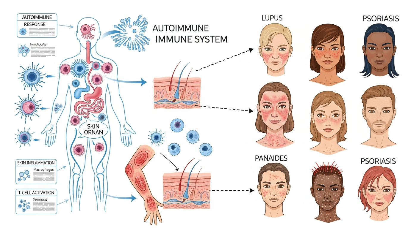
Lupus (systemic lupus erythematosus — SLE) produces several distinct skin manifestations, the most recognized being the malar rash — a butterfly-shaped redness across both cheeks and the bridge of the nose. This rash is flat or slightly raised, fixed (not transient like hives), photosensitive (worsens with sun exposure), and characteristically spares the nasolabial folds (the creases between the nose and mouth), which distinguishes it from rosacea and seborrheic dermatitis. However, not all lupus rashes are malar — discoid lupus erythematosus (DLE) produces scarring, coin-shaped plaques on the face, scalp, and ears. Subacute cutaneous lupus erythematosus (SCLE) causes widespread, symmetrical, non-scarring scaly red patches in sun-exposed areas. Drug-induced lupus from medications like hydralazine, minocycline, or procainamide can also cause skin manifestations. Any butterfly-shaped facial rash with joint pain, fatigue, oral ulcers, or abnormal blood tests should prompt evaluation for lupus.
Quick Medical Summary
The Malar Rash and Acute Cutaneous Lupus
The malar rash (malar butterfly rash) is the most iconic skin manifestation of systemic lupus erythematosus, occurring in approximately 50% of SLE patients. It appears as a fixed, erythematous (red) to violaceous (purple-pink) rash across both cheeks and the bridge of the nose, symmetrically, in the shape of a butterfly with wings spread. Key distinguishing features from the conditions it most resembles: (1) The malar rash spares the nasolabial folds (the smile lines from nose to mouth corners) — rosacea and seborrheic dermatitis affect these folds specifically. (2) It is fixed and persistent rather than transient — it doesn't fade within 24 hours as hives do. (3) It is exquisitely photosensitive — worsening within hours of sun exposure and improving with sun avoidance. (4) It is accompanied by systemic symptoms — joint pain, fatigue, oral ulcers, and abnormal laboratory findings (positive ANA, anti-dsDNA antibodies, low complement levels). On darker skin tones, the malar rash may appear as darker hyperpigmentation or violaceous discoloration rather than obvious redness, and photosensitivity may be easier to detect than the rash itself. Acute cutaneous lupus can also produce a widespread morbilliform rash resembling a drug eruption or viral rash — distinguishing features are the photosensitive distribution and positive lupus serology.
Discoid Lupus Erythematosus (DLE)
Discoid lupus erythematosus is a chronic, scarring form of cutaneous lupus that may occur with or without systemic lupus disease. DLE produces coin-shaped (discoid), well-defined, scaly plaques with follicular plugging (keratin filling and blocking hair follicles — seen as 'carpet tack' scale that reveals spiky projections when peeled off). Active DLE plaques are red and slightly raised at the periphery, with a pale, atrophic (thinned, scarred) center. Over time, the scarring becomes permanent — producing permanent alopecia (hair loss) in scalp involvement, depigmentation and scarring on the face and ears. The ears — particularly the conchal bowl (the hollow of the outer ear) and the ear canal rim — are a characteristic and often overlooked site for DLE. Facial DLE classically affects the malar areas, nose, and lips. About 5–10% of DLE patients develop systemic SLE; having DLE alone (without systemic disease) is not the same as having full SLE and does not carry the same visceral organ risks. DLE is treated with potent topical steroids, calcineurin inhibitors, and the antimalarial hydroxychloroquine (Plaquenil) — which reduces flare frequency, disease damage, and is protective against progression to SLE.
Other Lupus Skin Manifestations and When to Seek Evaluation
Subacute cutaneous lupus erythematosus (SCLE) produces two patterns: annular (ring-shaped, polycyclic) lesions or psoriasiform (scaling, plaque-like) lesions distributed symmetrically over sun-exposed areas — the upper back, shoulders, upper chest, and outer arms. Unlike DLE, SCLE does not scar. It is strongly associated with specific autoantibodies (anti-Ro/SSA) and with certain medications that can induce SCLE (hydrochlorothiazide, calcium channel blockers, proton pump inhibitors, terbinafine). Lupus panniculitis (lupus profundus) — a rare manifestation — causes painful, deep nodules beneath the skin on the face, arms, and buttocks, which heal with deep, permanent depressions. Livedo reticularis — a lacy, net-like purplish discoloration of the skin — occurs in lupus patients with antiphospholipid syndrome. Any patient with a facial butterfly rash, photosensitivity, mouth ulcers, hair loss, joint pain, fatigue, or unexplained blood cell count abnormalities should have lupus serology performed: antinuclear antibody (ANA), anti-double-stranded DNA (anti-dsDNA), anti-Smith, complement C3/C4 levels, and complete blood count. A strongly positive ANA (≥1:160) with compatible clinical features warrants rheumatology referral. The ACR/EULAR 2019 lupus classification criteria require ≥10 points across clinical and immunological domains for SLE classification.
Key Symptoms
- Fixed butterfly-shaped redness across cheeks and nose bridge, sparing nasolabial folds (malar rash)
- Rash worsening within hours of sun exposure and improving with sun avoidance (photosensitivity)
- Coin-shaped scarring plaques with follicular plugging on face, scalp, ears (DLE)
- Widespread scaly red patches symmetrically on sun-exposed upper body (SCLE)
- Malar rash alongside joint pain, fatigue, mouth ulcers, and hair loss
- Deep, painful nodules on face or arms healing with permanent indentations (lupus panniculitis)
Treatment Options
- Strict sun protection: SPF 50+ broad-spectrum sunscreen; UV-protective clothing; window film
- Hydroxychloroquine (Plaquenil): reduces flare frequency and protects against organ damage
- Topical corticosteroids for acute flares; intralesional steroids for DLE plaques
- Calcineurin inhibitors (tacrolimus) for facial lupus lesions avoiding steroid side effects
- Systemic immunosuppressants (azathioprine, mycophenolate) for extensive cutaneous involvement
- Rheumatology referral for suspected SLE; dermato-rheumatology collaboration for complex cases
When to See a Doctor Immediately
- Difficulty breathing or swallowing
- Swelling of the face, lips, or tongue
- High fever or severe chills
- Rapid spreading over a large body surface area
- Extreme pain, dizziness, or confusion
Frequently Asked Questions
Disclaimer
The medical information provided in this article is for educational purposes only and should not replace professional medical advice, diagnosis, or treatment. Always consult with a board-certified dermatologist or primary care physician regarding any severe or persistent skin conditions.